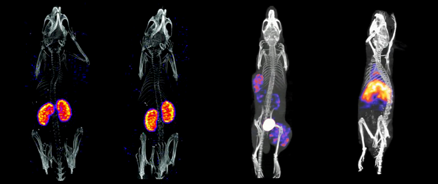
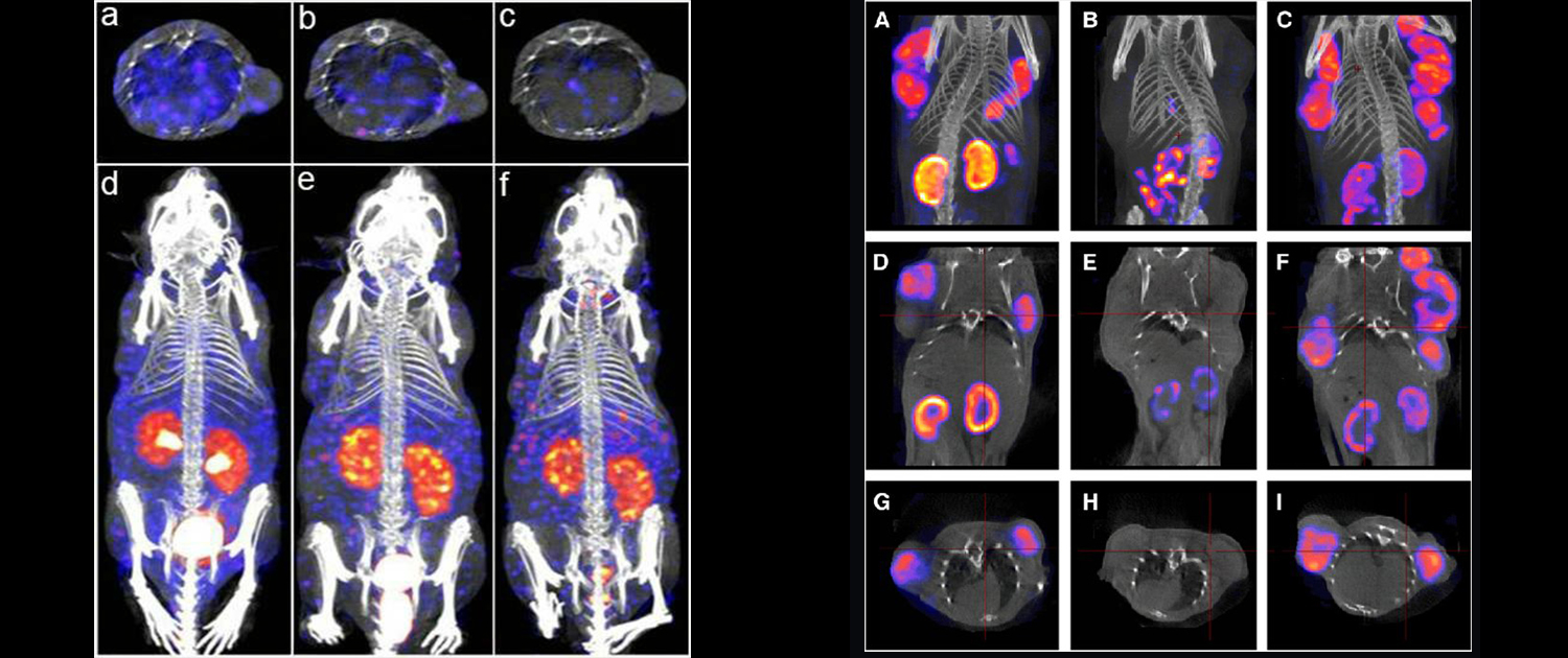
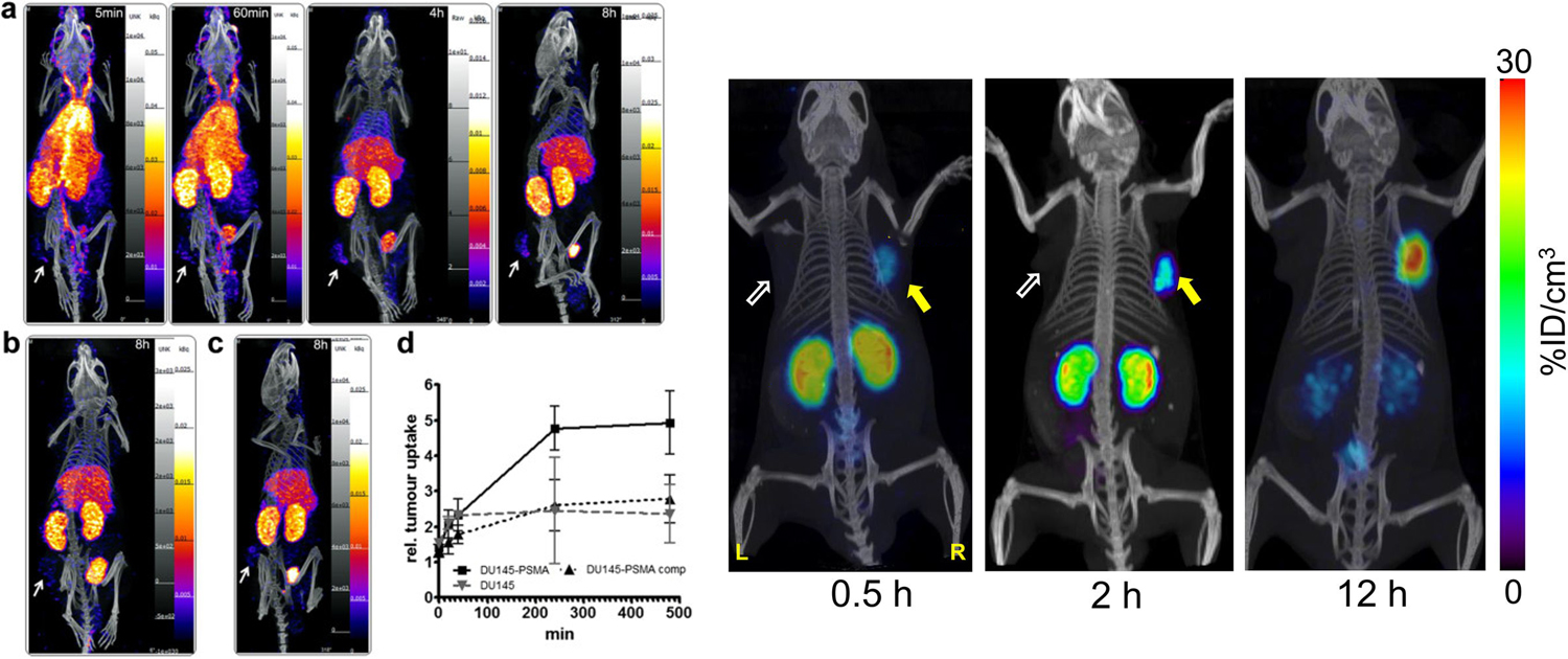
Micro-SPECT is a powerful imaging technique in preclinical research that utilizes gamma rays to provide high-resolution 3D images of biological processes in vivo. Similar to micro-PET, it uses radiotracers to visualize and quantify the distribution and uptake of specific molecules in tissues and organs. This makes it useful for studying a wide range of biological processes, including blood flow, metabolism, and receptor expression, among others. Micro-SPECT has numerous applications in preclinical research, including the development and evaluation of new drugs, disease models, and therapies.
Micro-SPECT has a broad range of applications in preclinical research, including studies on cancer, cardiovascular disease, and neurodegenerative diseases. It has also been used in drug development to evaluate the efficacy of new drugs and to identify potential drug targets. By providing detailed images of biological processes in vivo, micro-SPECT can help researchers gain insights into disease progression and treatment effectiveness, making it a valuable tool in preclinical research.
You can find information and description of the applications of Micro-SPECT in medical and preclinical research in the following.
Tumor imaging and characterization
Micro-SPECT is an imaging technique that allows for high-resolution and quantitative assessment of biological processes in small animals. One important application of micro-SPECT in preclinical research is the imaging and characterization of tumors. By using radiolabeled tracers, micro-SPECT can provide detailed information about tumor angiogenesis and blood flow, as well as pharmacokinetic parameters of potential therapeutic agents. This information can help researchers understand the underlying mechanisms of tumor growth and identify novel targets for therapeutic intervention. Additionally, micro-SPECT imaging can be used to monitor the response of tumors to therapy, providing valuable feedback on the efficacy of different treatments.
- Tumor imaging: Micro-SPECT can be used to visualize and quantify the distribution of radiotracers in tumors in small animals, providing valuable information on the size, location, and activity of the tumor. This information is important for assessing the efficacy of anti-cancer therapies and for developing new cancer treatments.
- Metastasis detection: Micro-SPECT can also be used to detect and quantify the spread of cancer cells to other parts of the body, allowing researchers to evaluate the effectiveness of anti-metastasis treatments and to develop new treatments to prevent cancer spread.
Cardiovascular research
Micro-SPECT is also a valuable tool for studying the cardiovascular system. It can be used to assess cardiac function and perfusion, providing important information about the mechanisms of heart disease. Additionally, micro-SPECT can be used to detect and monitor myocardial infarction, a common cause of heart failure. Finally, micro-SPECT can be used to study atherosclerosis, a chronic disease characterized by the buildup of plaque in the arteries. By using radiolabeled tracers, micro-SPECT can provide detailed information about the distribution of atherosclerotic lesions in small animals, allowing researchers to study the mechanisms of this disease.
- Cardiac function assessment: Micro-SPECT can be used to evaluate the function of the heart in small animals, providing valuable information on blood flow, perfusion, and cardiac contractility. This information is important for assessing the efficacy of cardiovascular drugs and for developing new treatments for heart disease.
- Myocardial infarction detection: Micro-SPECT can also be used to detect and quantify the extent of myocardial infarction in small animals, allowing researchers to evaluate the effectiveness of treatments for heart attacks and to develop new treatments to prevent heart damage.
Drug development and pharmacology
Micro-SPECT is a powerful tool for studying drug development and pharmacology. It can be used to assess the metabolism and distribution of potential therapeutic agents, providing important information about their pharmacokinetic properties. Additionally, micro-SPECT can be used to assess pharmacodynamics or the effects of drugs on biological processes in vivo. Finally, micro-SPECT can be used to evaluate the efficacy of new drug candidates, providing important information about their potential as therapeutic agents.
- Pharmacokinetic studies: Micro-SPECT can be used to track the distribution and concentration of radiotracers in small animals over time, providing valuable information on the pharmacokinetics of drugs. This information is important for optimizing drug dosing and for evaluating the efficacy and safety of new drugs.
- Drug target validation: Micro-SPECT can also be used to visualize and quantify the distribution of radiotracers that bind to specific drug targets in small animals, providing valuable information on the role of the drug target in disease and in response to therapies.
Immunology and targeted therapies
Micro-SPECT is a valuable tool for molecular imaging and the development of targeted therapies. It can be used to image and quantify specific molecular targets in vivo, providing important information about the expression and distribution of these targets. This can be used to develop targeted therapies, such as targeted radiotherapy, which uses radiolabeled agents to selectively kill cancer cells. Finally, micro-SPECT can be used to assess the efficacy of targeted therapies.
- Inflammation imaging: Micro-SPECT can be used to visualize and quantify the distribution of radiotracers in inflamed tissues in small animals, providing valuable information on the location and activity of inflammatory cells. This information is important for evaluating the efficacy of anti-inflammatory drugs and for developing new treatments for inflammatory diseases.
- Immune cell tracking: Micro-SPECT can also be used to track the distribution and concentration of immune cells in small animals, providing valuable information on the role of the immune system in disease and in response to therapies.
Neuroimaging and neuroscience research
Another important application of micro-SPECT in preclinical research is in the field of neuroimaging and neuroscience. Micro-SPECT can be used to study the distribution and function of neurotransmitter receptors in the brain, providing insight into the mechanisms of neuropsychiatric disorders. Additionally, micro-SPECT can be used to map neuronal pathways and assess the connectivity of different brain regions. This can help researchers understand the neural basis of behavior and cognition. Micro-SPECT is also a useful tool for studying neurodegenerative diseases, such as Alzheimer’s and Parkinson’s, as it can provide detailed information about the distribution of pathology in the brain.
- Brain imaging: Micro-SPECT can be used to visualize and quantify the distribution of radiotracers in the brain of small animals, providing valuable information on the structure and function of the brain. This information is important for studying brain development, assessing the efficacy of drugs for neurological disorders, and for developing new treatments for brain diseases.
- Neurotransmitter mapping: Micro-SPECT can also be used to map the distribution of neurotransmitters in the brain of small animals, providing valuable information on the role of neurotransmitters in normal brain function and in neurological disorders.
What can we do for you?
We offer a wide range of preclinical imaging services, including micro-PET, micro-SPECT, micro-CT, and optical imaging. Our team of experienced specialists is dedicated to providing high-quality and reliable image analysis services to help researchers in the preclinical research field. Researchers can register their research projects on our website and choose the services they need or set up a free virtual meeting with our specialists to discuss and get guidance about the available related services that we can perform for them.
Our image analysis services are customizable and can be tailored to fit the specific needs of each research project. With our state-of-the-art equipment and cutting-edge image analysis techniques, we aim to help researchers obtain the most accurate and informative results from their preclinical imaging studies.
To register your research project with our company, please register your project through the “Contact Us” page on our website and fill out the required fields, including your name, email address, and project details.
We will review your submission and contact you as soon as possible to schedule a free virtual meeting with our imaging specialists, who can provide guidance and support for your research.


