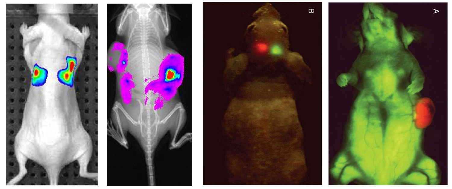
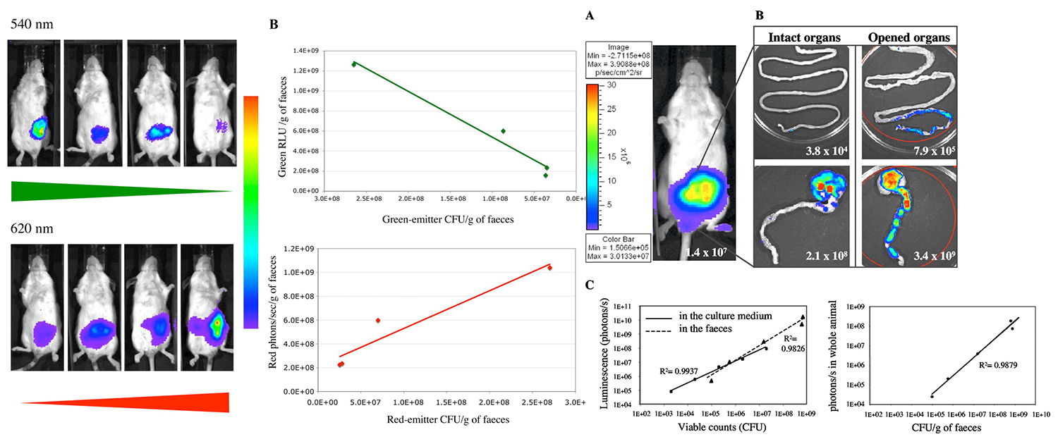
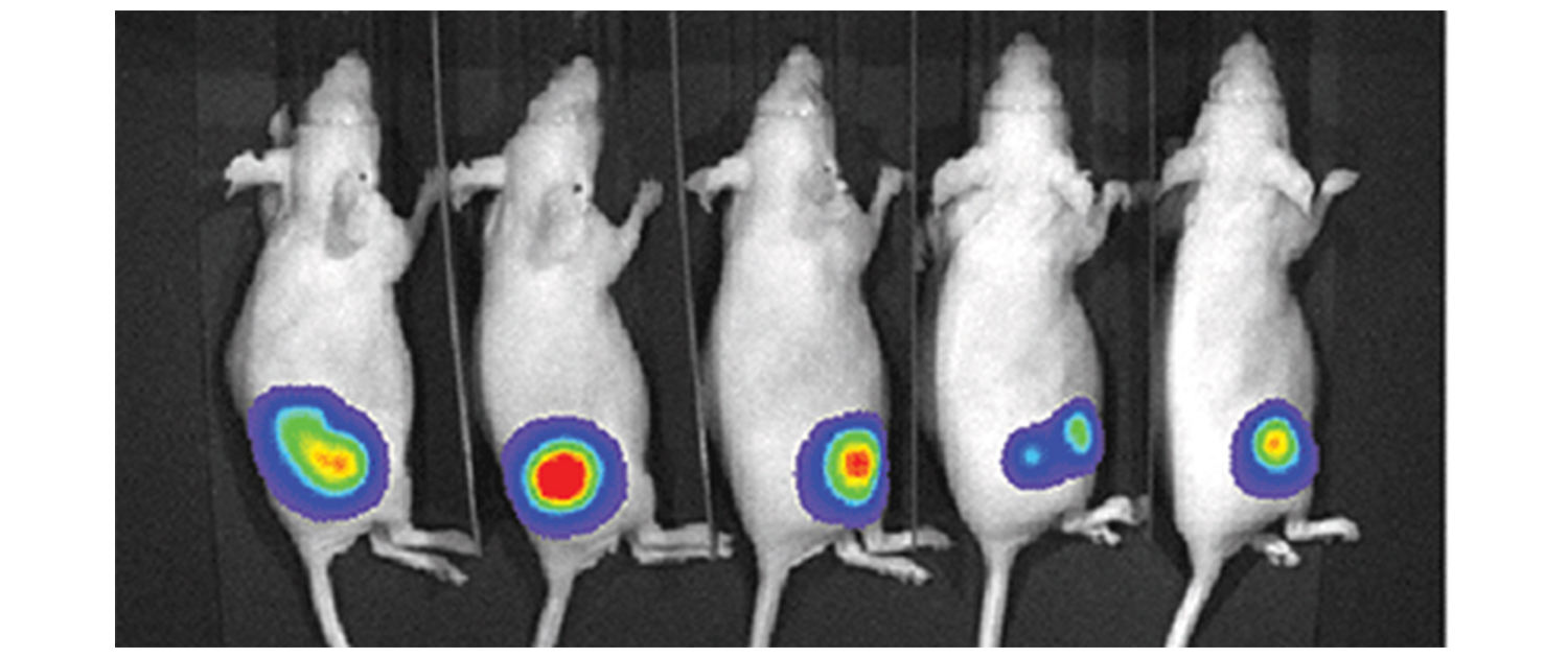
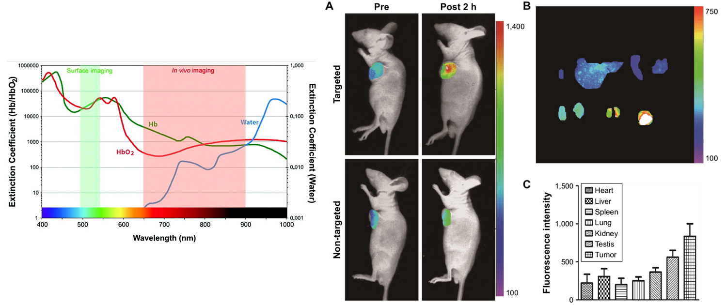
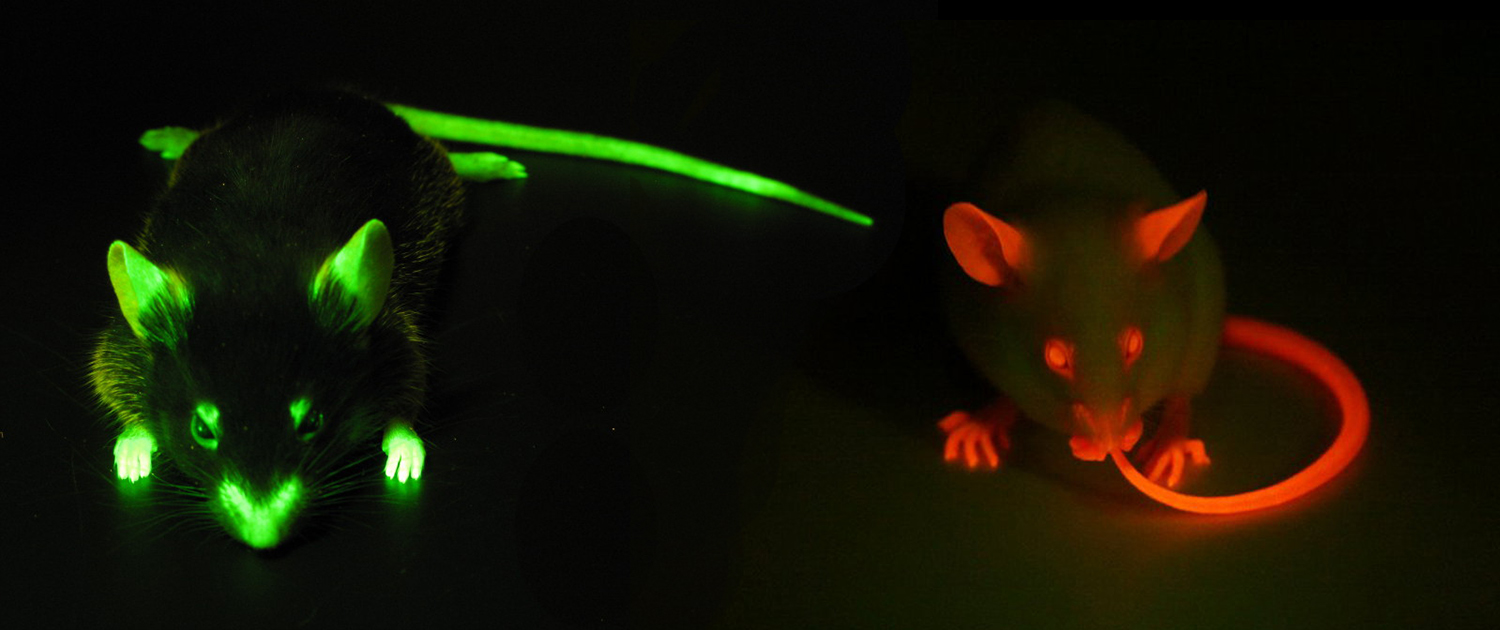
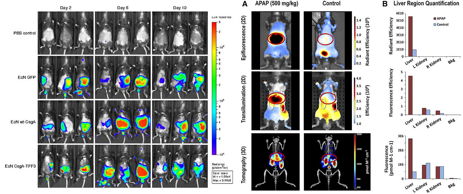
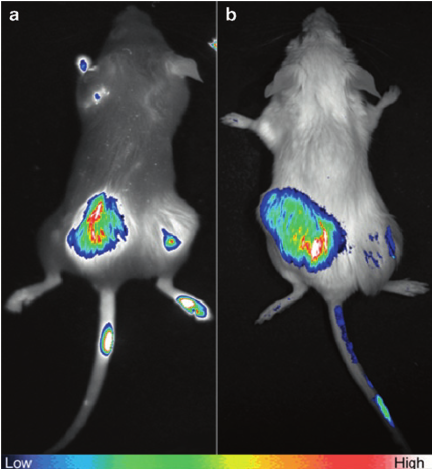
Optical imaging, including fluorescent imaging, has become a popular modality in preclinical research due to its ability to provide real-time information on biological processes in vivo. The use of fluorescent dyes and probes enables researchers to selectively label and visualize specific cells, proteins, and molecular interactions in real-time. Optical imaging techniques such as fluorescence microscopy, bioluminescence, and optoacoustic imaging have all been used to investigate a wide range of biological processes in vivo, including tumor biology, drug distribution, and cellular interactions. One major benefit of optical imaging is its ability to provide high-resolution images of biological processes in vivo.
This is particularly useful for investigating complex biological systems, such as the brain, where traditional imaging techniques are limited in their ability to provide detailed information. By using fluorescent dyes and probes, researchers can selectively label and visualize specific cells, proteins, and molecular interactions, providing detailed information about their behavior, interactions, and spatial distribution within tissues and organs. This information can be used to better understand the underlying mechanisms of disease and to identify potential therapeutic targets.
You can find information and description of the applications of optical imaging in preclinical research in the following.
Tumor imaging and characterization
Optical imaging has become an important tool for the visualization and characterization of tumors in preclinical research. It can be used for early cancer detection by visualizing biomarkers of tumor development and angiogenesis. Optical imaging can also be used to visualize the tumor microenvironment, including the vasculature, stromal cells, and immune cells. Finally, optical imaging can be used to monitor tumor response to therapy, providing valuable information about the efficacy of different treatments.
Neuroimaging and neuroscience research
Optical imaging is a powerful tool for the study of the brain and the neural processes that underlie behavior and cognition. It can be used for in vivo imaging of brain function, providing detailed information about changes in blood flow, oxygenation, and neurotransmitter levels. Optical imaging can also be used for the study of neurodegenerative diseases, providing insight into the pathophysiology of diseases such as Alzheimer’s and Parkinson’s. Finally, optical imaging can be used to visualize neuronal activity, providing valuable information about the patterns of activation associated with different behaviors.
Cardiovascular research
Optical imaging is a valuable tool for the study of the cardiovascular system in preclinical research. It can be used for the study of vascular function, providing information about blood flow, endothelial function, and angiogenesis. Optical imaging can also be used to detect and monitor myocardial infarction, a common cause of heart failure. Finally, optical imaging can be used to study atherosclerosis, providing valuable information about the distribution of atherosclerotic lesions in the vasculature and the mechanisms underlying the disease.
Drug development and pharmacology
Optical imaging is an important tool for the study of drug development and pharmacology in preclinical research. It can be used for the study of drug pharmacokinetics, providing information about drug distribution and metabolism in vivo. Optical imaging can also be used to assess drug efficacy, providing information about the effects of drugs on biological processes. Finally, optical imaging can be used for drug discovery, providing a platform for the high-throughput screening of potential drug candidates.
Molecular imaging and targeted therapies
Optical imaging is a valuable tool for the study of molecular imaging and targeted therapies in preclinical research. It can be used for the visualization of specific molecular targets in vivo, providing important information about the expression and distribution of these targets. This can be used to develop targeted therapies, such as antibody-based therapies, which selectively target cancer cells. Additionally, optical imaging can be used to study gene expression, providing a non-invasive tool for monitoring the activity of gene therapy vectors in vivo. Overall, optical imaging is a powerful tool for the study of molecular mechanisms in preclinical research, and has the potential to lead to the development of new and effective treatments for a wide range of diseases.
What can we do for you?
We offer a wide range of preclinical imaging services, including micro-PET, micro-SPECT, micro-CT, and optical imaging. Our team of experienced specialists is dedicated to providing high-quality and reliable image analysis services to help researchers in the preclinical research field. Researchers can register their research projects on our website and choose the services they need or set up a free virtual meeting with our specialists to discuss and get guidance about the available related services that we can perform for them.
Our image analysis services are customizable and can be tailored to fit the specific needs of each research project. With our state-of-the-art equipment and cutting-edge image analysis techniques, we aim to help researchers obtain the most accurate and informative results from their preclinical imaging studies.
To register your research project with our company, please register your project through the “Contact Us” page on our website and fill out the required fields, including your name, email address, and project details.
We will review your submission and contact you as soon as possible to schedule a free virtual meeting with our imaging specialists, who can provide guidance and support for your research.

