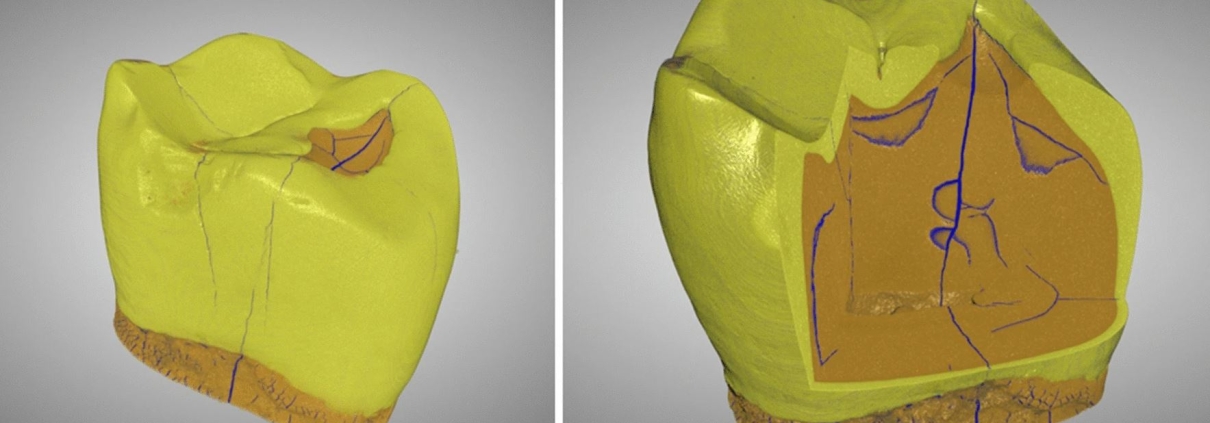
Micro CT Applications in Dentistry
An insightful article by Dr. Akshima Sahi, BDS, discusses the diverse applications of microcomputed tomography (Micro-CT) in dentistry. This cutting-edge technology is revolutionizing dental research and treatment with its ability to produce high-resolution, non-destructive 3D images of biological structures like teeth.
“Micro CT has varied uses in dentistry ranging from dental research to treatment. Micro CT produces high-quality images from the outer to the innermost structure of the tooth and surrounding structures. As micro CT creates non-destructive images, the same image can be viewed multiple times and the image remains incessantly for biological and mechanical testing.”
Key highlights from the article:
- Accurate measurement of enamel and inner tooth structures, including dentin and pulp chambers.
- Enhanced understanding of root canal morphology and improved root canal treatment outcomes.
- Analysis of craniofacial skeletal development and trabecular bone structure.
- Evaluation of dental implants for stabilization and osseointegration.
- Applications in tissue engineering and finite element modeling (FEM) for biomechanical studies.
At KARA, we specialize in micro-CT, image analysis, and preclinical imaging solutions, offering advanced image analysis to support research and innovation in dentistry and beyond.
Read the full article here: news-medical.net
Let us know how Micro-CT can help with your research or clinical needs!
*Image from: Scientific Reports (Sci Rep) ISSN 2045-2322 (online)

 nature.com
 sagepub.com
Leave a Reply
Want to join the discussion?Feel free to contribute!