Image Analysis

Image analysis is a critical component of clinical and preclinical imaging research, providing a means of quantifying and interpreting the data generated by imaging modalities such as PET, SPECT, and CT. In clinical imaging, image analysis is used to aid in diagnosis and treatment planning. For example, in oncology, image analysis can be used to assess the response to chemotherapy and radiation therapy, providing a means of monitoring the effectiveness of treatment and making adjustments as necessary.

In cardiology, image analysis can be used to evaluate blood flow and cardiac function, enabling the diagnosis and treatment of heart disease. In preclinical imaging research, image analysis plays a crucial role in the development of new treatments and therapies. Image analysis algorithms can be used to quantify the effects of drug treatments or surgical procedures on biological processes such as blood flow and metabolic activity. By providing a means of objectively assessing treatment efficacy, image analysis helps to optimize treatment protocols and improve patient outcomes.

You can find information and description of the basics and applications of image analysis in medical and preclinical research in the following.

Image Analysis in Micro-PET Imaging

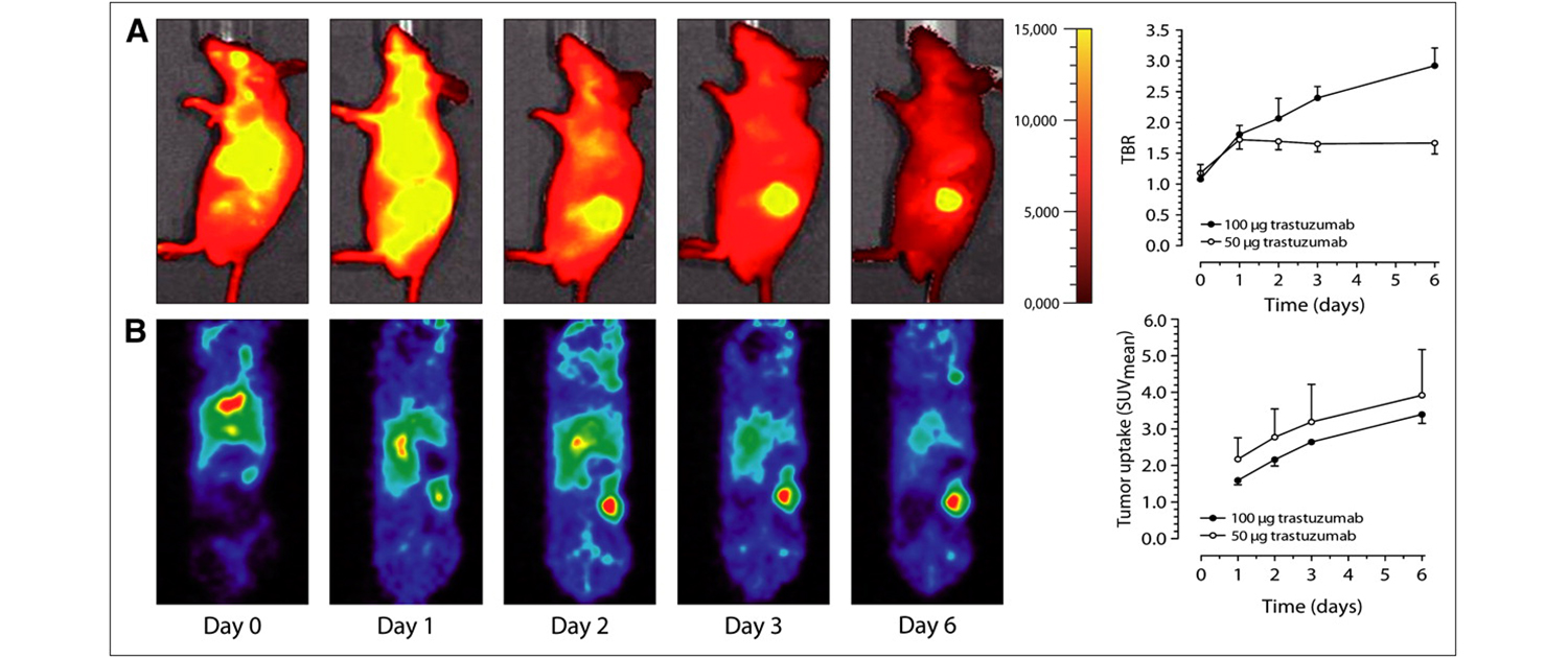
One of the key applications of image analysis in preclinical micro-PET is the quantification of radiotracer uptake in different regions of interest. This technique helps in determining the amount of radiotracer uptake in different tissues and organs of the animal model, and can provide valuable information about the distribution and pharmacokinetics of the radiotracer.

Micro-PET imaging combined with image analysis can be used to measure tumor volume in preclinical research models. This technique is particularly useful in evaluating the effectiveness of cancer therapies, as it allows researchers to track changes in tumor size over time.

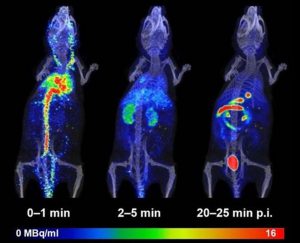
Dynamic PET imaging is a technique that involves the continuous acquisition of PET images over time, which can provide important information about the kinetics of radiotracer uptake and distribution in preclinical research models. Image analysis of dynamic PET data can provide quantitative information about tissue perfusion, blood flow, and other physiological parameters.

Micro-PET imaging can also be used to study brain function in preclinical research models, and image analysis can be used to identify specific regions of the brain that are activated in response to various stimuli. This technique is useful in studying a range of neurological and psychiatric disorders and can provide valuable insights into the underlying mechanisms of these conditions.

In addition to quantifying radiotracer uptake in different tissues, image analysis can also be used to model the pharmacokinetics of radiotracers in preclinical research models. This information can be used to optimize drug dosing and evaluate the efficacy of new drugs in development.

Image Analysis in Micro-SPECT Imaging

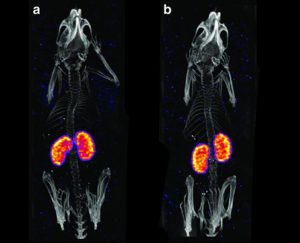
Similar to micro-PET, micro-SPECT imaging combined with image analysis can be used to quantify radiotracer uptake in different tissues and organs of preclinical research models. This technique is particularly useful in studying the distribution and pharmacokinetics of radiotracers.

Micro-SPECT imaging is commonly used in preclinical research to study cardiac function, and image analysis can be used to assess a range of cardiac parameters such as myocardial perfusion, left ventricular function, and cardiac output. This technique can be used to evaluate the effectiveness of cardiac therapies and drug candidates.

Micro-SPECT imaging combined with image analysis can also be used to evaluate tumor targeting and measure tumor volume in preclinical research models. This technique can be used to track changes in tumor size over time and evaluate the effectiveness of cancer therapies.

Micro-SPECT imaging can be used to study bone and joint function in preclinical research models, and image analysis can be used to evaluate bone mineral density, bone metabolism, and joint function. This technique is useful in studying a range of musculoskeletal disorders and can provide valuable insights into disease progression and treatment efficacy.

Micro-SPECT imaging can be used to study inflammation and infection in preclinical research models, and image analysis can be used to evaluate the extent of inflammation and the distribution of infectious agents. This technique can be used to evaluate the efficacy of anti-inflammatory drugs and antimicrobial agents.

Image Analysis in Micro-CT Imaging

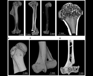
Micro-CT imaging is a powerful tool for studying bone microarchitecture in preclinical research, dental research, and industry. Image analysis techniques can be used to extract quantitative information from the images, such as bone mineral density, bone volume fraction, and trabecular thickness. These parameters can be used to assess bone quality and to monitor the effects of interventions on bone health.

Micro-CT imaging can also be used in tissue engineering research for the evaluation of scaffold architecture and pore size distribution. Image analysis can be used to extract important parameters such as porosity, pore size, and interconnectivity to assess the suitability of a scaffold for a particular application.

In preclinical research, micro-CT imaging can be used to assess angiogenesis in vivo. Image analysis can be used to calculate vascular parameters such as vessel volume, vessel surface area, and vessel length, providing important information on the development of new blood vessels.

Micro-CT imaging is also a valuable tool in preclinical tumor imaging, allowing for the monitoring of tumor growth and the assessment of treatment efficacy. Image analysis can be used to extract tumor volume and vascularization information, providing important information on the development of tumors.

Micro-CT imaging is widely used in industry for non-destructive testing and analysis. Image analysis techniques can be used to extract quantitative information from the images, such as porosity, crack density, and fiber orientation, to assess the quality of industrial products such as composites, ceramics, and metals. The use of micro-CT imaging and image analysis in industry allows for the optimization of production processes and the improvement of product performance.

Image Analysis in Dental Micro-CT Imaging

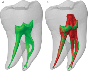
Micro-CT can be used to image and analyze the complex anatomy of the root canal system, providing detailed information on the shape and size of the canal. Image analysis can be used to identify the presence of root canal fillings, detect fractures or perforations, and evaluate the effectiveness of endodontic treatment.

Micro-CT is used to create a 3D model of the jawbone and teeth, allowing for accurate placement of dental implants. Image analysis can be used to measure bone density and quality, identify potential implant sites, and simulate the placement of the implant in the model.

Micro-CT can be used to examine the growth and development of teeth, providing high-resolution 3D images of the internal structure of the tooth. Image analysis can help to quantify and visualize different aspects of tooth development, such as the growth of enamel and dentin layers.

Micro-CT can be used to evaluate the properties of dental materials such as fillings, crowns, and implants. Image analysis can provide information on material wear, fracture, and degradation over time, allowing for the optimization of these materials and improving their performance.

Micro-CT can be used to visualize and quantify bone loss due to periodontal disease. Image analysis can help to assess the degree of bone loss and to identify areas of active disease, providing important information for the diagnosis and treatment of periodontal disease.

Image Analysis in Optical Imaging

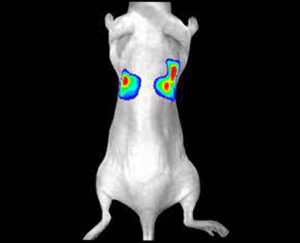
Optical imaging is frequently used to study tumor biology in preclinical research models, and image analysis can be used to evaluate tumor microenvironment parameters such as blood flow, vascular permeability, and oxygenation. This technique is useful in evaluating the effectiveness of anti-cancer therapies and identifying novel therapeutic targets.

Optical imaging can be used to study cardiovascular function in preclinical research models, and image analysis can be used to assess cardiac function, vascular permeability, and blood flow parameters. This technique is useful in evaluating the efficacy of cardiovascular therapies and identifying new drug targets.

Optical imaging can also be used in preclinical research to study the immune system, and image analysis can be used to evaluate immune cell infiltration, cytokine expression, and immune cell activation. This technique can be used to evaluate the efficacy of immunotherapies and identify new therapeutic targets.

Optical imaging can be used to study brain function in preclinical research models, and image analysis can be used to assess brain perfusion, neuronal activity, and other brain parameters. This technique is useful in evaluating the efficacy of neurological therapies and identifying new drug targets.

Optical imaging can be used to study infectious diseases in preclinical research models, and image analysis can be used to evaluate the extent of infection and the distribution of infectious agents. This technique is useful in evaluating the efficacy of antimicrobial agents and identifying new therapeutic targets.

The same image analysis techniques can be applied to both clinical and preclinical images, allowing for a seamless transition between research modalities. Moreover, image analysis has additional applications in clinical research, beyond the evaluation of disease progression or response to therapy. For example, image analysis can be used for radiation treatment planning to optimize the radiation dose and minimize side effects.

Image analysis can also be used to evaluate the efficacy of medical devices or surgical procedures. Additionally, image analysis can be used in radiomics, which involves the high-throughput extraction of large amounts of data from medical images, allowing for the identification of imaging biomarkers that can help in patient diagnosis and prognosis. Overall, image analysis is a powerful tool in both preclinical and clinical research, with a wide range of applications beyond disease evaluation.

WHAT CAN WE DO FOR YOU?

At our company, we provide a wide range of image analysis services for both preclinical and clinical imaging research. Our team of experts has extensive experience in conducting image analysis for various modalities, including PET, SPECT, CT, and optical imaging. We offer a comprehensive set of services, such as image registration, segmentation, quantification, and statistical analysis. We understand the importance of accurate and precise image analysis in the success of research projects, and we use state-of-the-art software and techniques to deliver reliable and reproducible results.

If you are a researcher looking for image analysis services, you can register your research project with us, and our team will work with you to develop a customized plan that suits your research needs. We also offer a free consultancy virtual meeting with our specialists to discuss your research project and provide guidance about the available image analysis services that we can perform for you. Our team is committed to providing high-quality services to our clients and helping them achieve their research goals.

To register your research project with our company, please register your project through the “Contact Us” page on our website and fill out the required fields, including your name, email address, and project details.

We will review your submission and contact you as soon as possible to schedule a free virtual meeting with our imaging specialists, who can provide guidance and support for your research.