Micro-PET
Micro-SPECT
Micro-CT
Optical Imaging
Preclinical imaging refers to the use of imaging techniques to visualize and study biological processes in animals before human clinical trials. Preclinical imaging is a crucial tool in medical and pharmaceutical research as it allows researchers to non-invasively study the effects of drugs and diseases on living systems. It helps in developing new treatments and improving existing ones. Preclinical imaging also plays a significant role in understanding the underlying mechanisms of diseases, identifying biomarkers, and predicting treatment outcomes.
Micro-PET, Micro-SPECT, Micro-CT, and optical imaging are some of the most widely used preclinical imaging modalities. Micro-PET and Micro-SPECT are highly sensitive and allow for the non-invasive imaging of biochemical processes in vivo.
These techniques are commonly used to study molecular pathways and metabolic processes in animals, aiding in the identification and validation of drug targets. Micro-CT imaging, on the other hand, provides high-resolution three-dimensional images of biological structures, making it an invaluable tool for anatomical studies and tissue engineering research. Optical imaging is a non-invasive imaging technique that uses light to visualize biological processes, allowing for real-time monitoring of biological activities in animals. It is often used to study cellular and molecular processes, such as gene expression and protein interactions, and can aid in the development of targeted therapies.
You can find information and description of applications of some preclinical imaging systems in the following.
Micro-PET
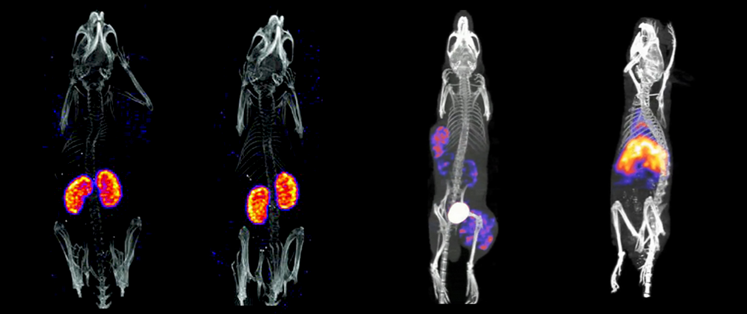
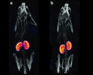
Micro-PET, or micro-Positron Emission Tomography, is an imaging technique that uses radiolabeled molecules to track biological processes in animals. In this technique, animals are injected with a radiotracer that emits positrons. When the positrons collide with electrons in the tissue, they release gamma rays, which are detected by the scanner to create a 3D image of the body. Micro-PET has applications in drug development, cancer research, and neurology. For example, micro-PET can be used to study drug metabolism and distribution in vivo, to track the progression of tumors, and to study neurodegenerative diseases.
Micro-SPECT
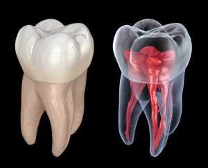
Micro-SPECT, or micro-Single Photon Emission Computed Tomography, is another imaging technique that uses radiolabeled molecules to track biological processes. In this technique, animals are injected with a radiotracer that emits gamma rays. The gamma rays are detected by a scanner to create a 3D image of the body. Micro-SPECT has applications in the study of cardiovascular diseases, cancer, and neurological disorders. For example, micro-SPECT can be used to study blood flow in the heart, to track the spread of cancer, and to study brain activity.
Micro-CT
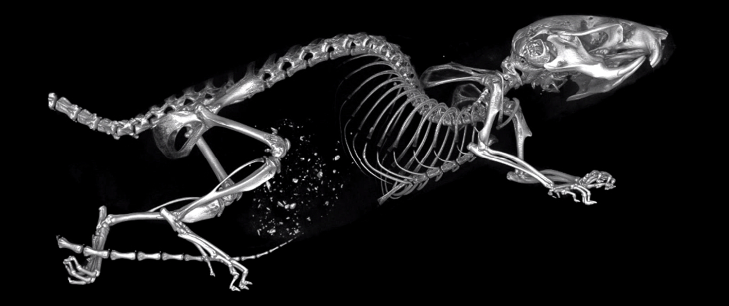
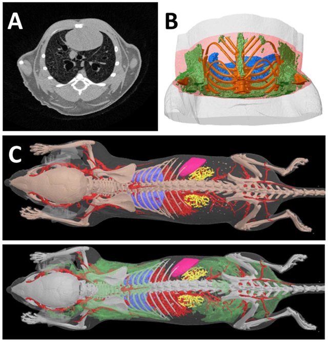
Micro-CT, or micro-Computed Tomography, is an imaging system that provides high-resolution 3D images of biological structures. This system uses X-rays to create cross-sectional images of the body, which are then reconstructed into a 3D image. Micro-CT has applications in the study of anatomical structures, bone density, and tissue engineering. For example, micro-CT can be used to study bone structure and density, to analyze the microarchitecture of tissues, and to study the growth of tumors.
Optical Imaging
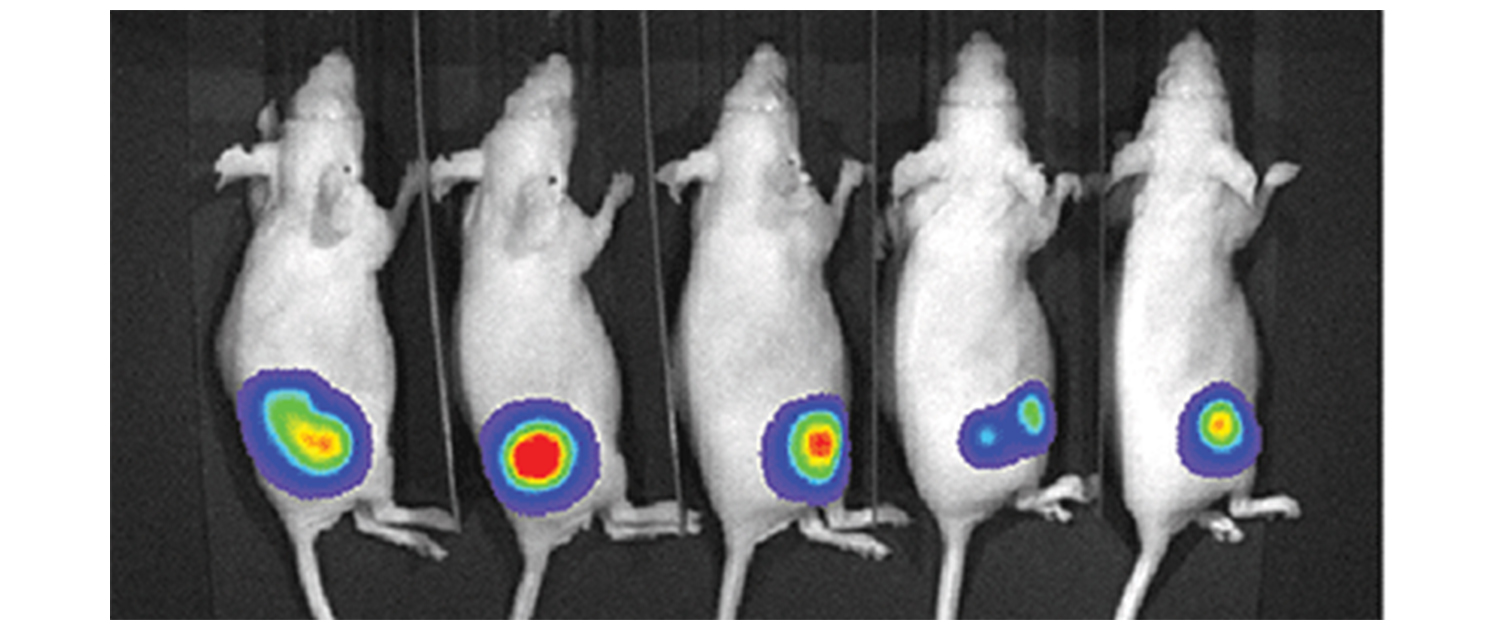
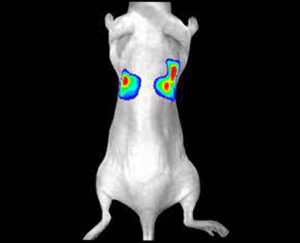
Optical imaging is a non-invasive imaging technique that uses light to visualize biological processes. This system uses fluorescent probes to track biological processes in vivo, which emit light when excited by a specific wavelength of light. Optical imaging has applications in the study of cellular and molecular processes, such as gene expression, protein interactions, and cancer research. For example, optical imaging can be used to study the distribution of specific cells, to track the expression of genes, and to monitor the activity of enzymes.
What can we do for you?
We offer a wide range of preclinical imaging services, including micro-PET, micro-SPECT, micro-CT, and optical imaging. Our team of experienced specialists is dedicated to providing high-quality and reliable image analysis services to help researchers in the preclinical research field. Researchers can register their research projects on our website and choose the services they need or set up a free virtual meeting with our specialists to discuss and get guidance about the available related services that we can perform for them.
Our image analysis services are customizable and can be tailored to fit the specific needs of each research project. With our state-of-the-art equipment and cutting-edge image analysis techniques, we aim to help researchers obtain the most accurate and informative results from their preclinical imaging studies.
To register your research project with our company, please register your project through the “Contact Us” page on our website and fill out the required fields, including your name, email address, and project details.
We will review your submission and contact you as soon as possible to schedule a free virtual meeting with our imaging specialists, who can provide guidance and support for your research.


