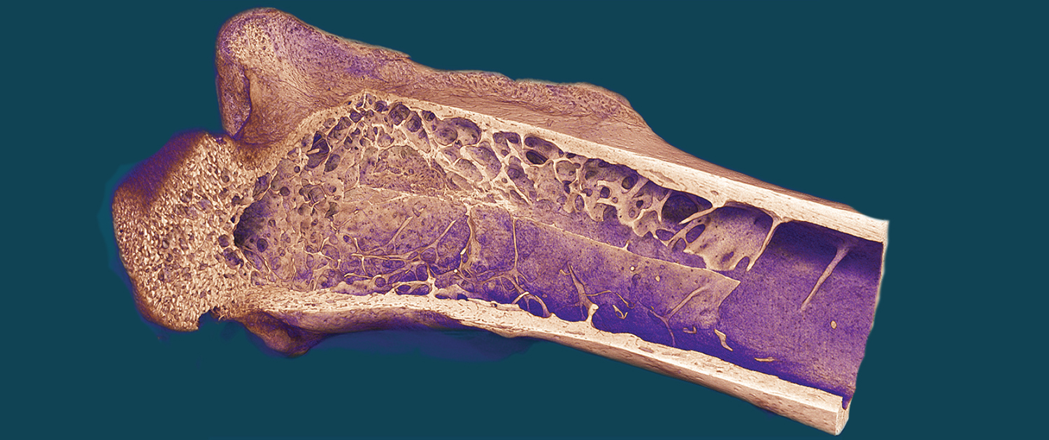
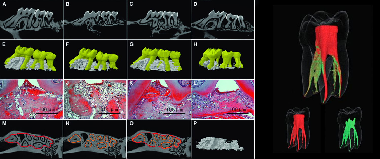
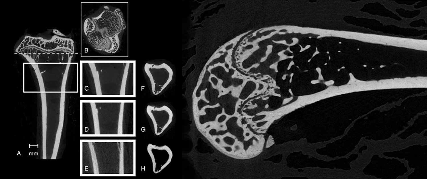
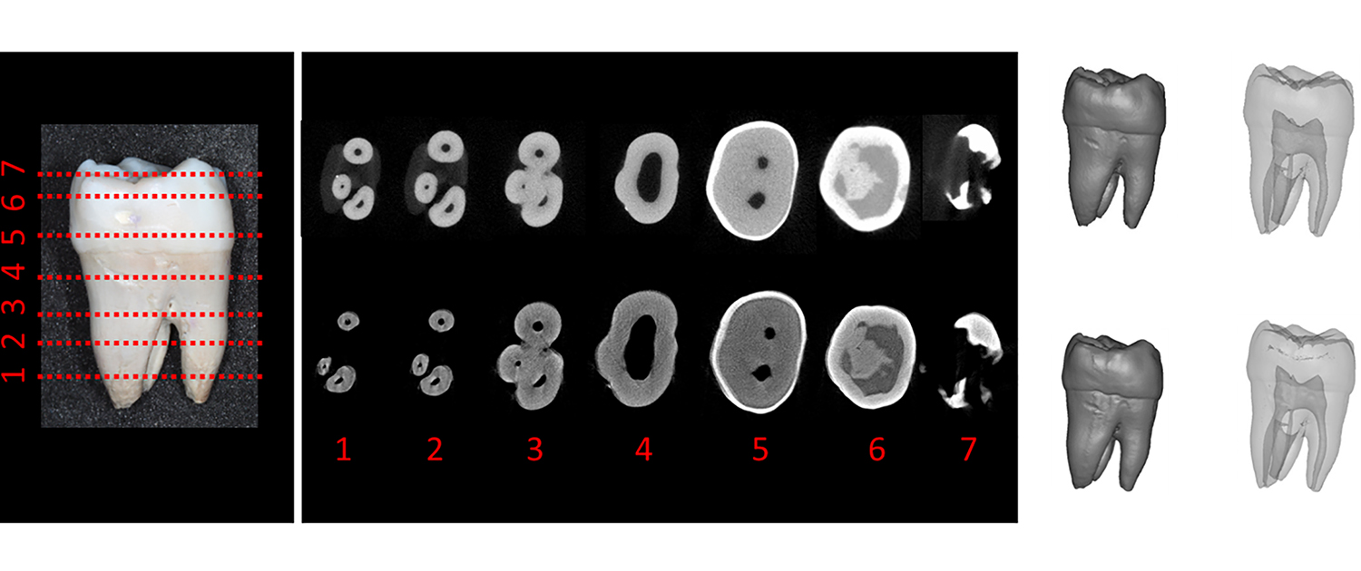
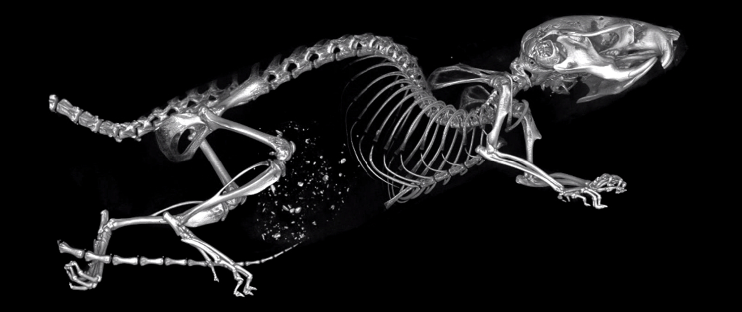
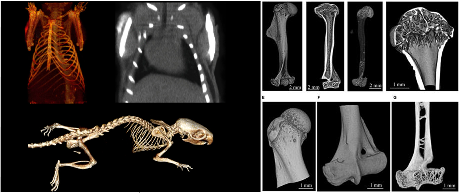
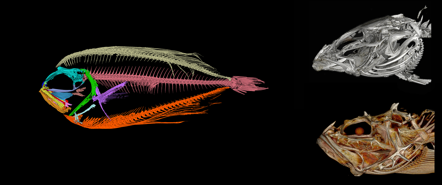
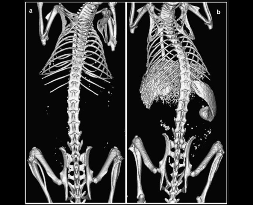
Micro-Computed Tomography (micro-CT) is a non-invasive imaging technology that is widely used in both preclinical and clinical research. In preclinical research, micro-CT is employed to visualize internal anatomy and assess bone density, tissue composition, and microarchitecture, and monitor disease progression in animal models. It is also used to evaluate the efficacy of therapeutic interventions and assess the safety of new medical devices and drugs. Micro CT scans provide high-resolution, three-dimensional images of internal anatomy, which can reveal intricate details about the biological structure of tissues and organs.
In clinical research, micro-CT is used to examine bones and joints, detect early signs of osteoporosis and assess the therapeutic response to treatments. It is also used in dental research to evaluate the mineral density and structure of teeth, and in cardiology to visualize the coronary arteries and assess the distribution of plaque in the arterial walls. The ability to visualize internal anatomy in such detail provides valuable information for researchers and clinicians, allowing them to make more informed decisions about patient care. Additionally, micro-CT is also useful in forensic medicine, allowing the examination of bones to determine the cause of injury or death.
You can find information and description of the applications of Micro-CT imaging in the following.
Micro-CT in Preclinical Imaging
Organ Morphology Evaluation
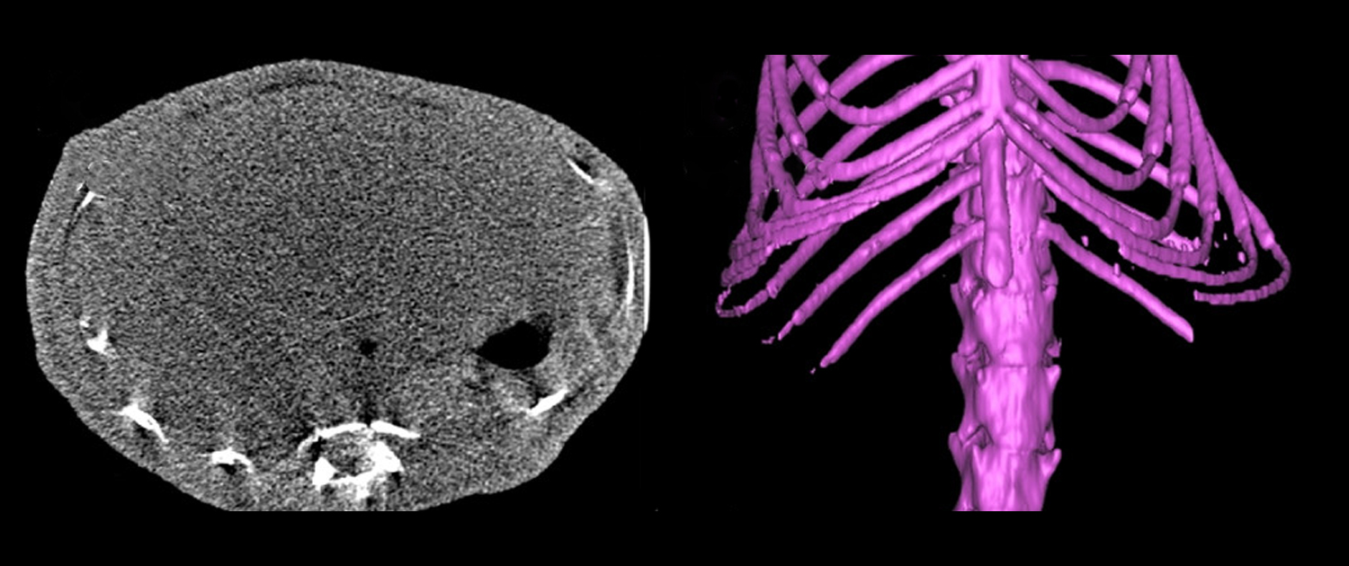
Micro CT provides researchers with a non-invasive method for assessing organ morphology in small animal models. The 3D images generated by the system allow for a detailed evaluation of the structure and function of various organs, including the heart, liver, and lungs.
Cancer research
- Tumor imaging: Micro-CT can be used to visualize tumors in preclinical models, allowing researchers to track tumor growth and response to treatment. This information can be used to develop new cancer therapies.
- Angiogenesis analysis: Micro-CT can be used to study the formation of new blood vessels in tumors, a process known as angiogenesis. This is important for understanding tumor growth and for developing new anti-angiogenic therapies.
- Metastasis detection: Micro-CT can also be used to detect metastatic tumors, allowing researchers to study the process of cancer spread and develop new therapies to prevent it.
Disease Progression Study
Micro CT is used to study the progression of various diseases and disorders in small animal models. The system’s ability to generate high-resolution, 3D images provides researchers with a comprehensive understanding of the effects of these diseases on the anatomy and function of the body.
Lung Imaging
- Pulmonary function assessment: Micro-CT can be used to assess lung function in preclinical models, allowing researchers to study lung diseases such as asthma and chronic obstructive pulmonary disease (COPD).
- Airway analysis: Micro-CT can also be used to analyze airway structure and function, which is useful in understanding the underlying mechanisms of lung diseases and for developing new treatments.
- Imaging of pulmonary diseases: Micro-CT can be used to visualize lung diseases such as fibrosis and emphysema in preclinical models, allowing researchers to study the progression of these diseases and develop new therapies.
Cardiovascular research
- Vascular imaging: Micro-CT can be used to visualize blood vessels in preclinical models, allowing researchers to study cardiovascular disease and develop new therapies.
- Cardiac function assessment: Micro-CT can also be used to assess cardiac function in preclinical models, allowing researchers to study conditions such as heart failure and develop new treatments.
- Atherosclerosis analysis: Micro-CT can be used to study the development of atherosclerosis, a condition where plaque builds up in arteries, leading to cardiovascular disease.
Heart Structure Visualization
Micro CT provides researchers with a non-invasive method for visualizing the internal structure of the heart and blood vessels in small animal models. This information is valuable for the study of cardiovascular disease and the development of new treatments.
Neurological research
- Brain imaging: Micro-CT can be used to visualize the structure of the brain in preclinical models, allowing researchers to study neurological conditions such as Alzheimer’s disease and develop new treatments.
- Cranial nerve analysis: Micro-CT can also be used to study cranial nerves, which play an important role in various neurological functions.
- Neurovascular analysis: Micro-CT can be used to study the interaction between neurons and blood vessels, which is important for understanding neurological diseases and developing new therapies.
Micro-CT in Bone Studies
Bone Density Analysis
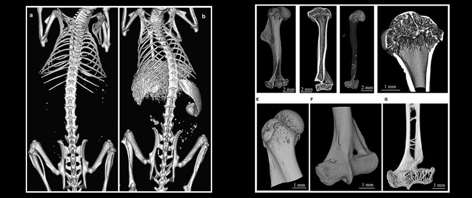
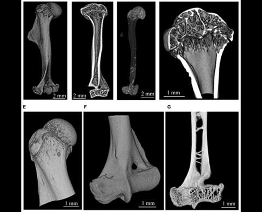
Micro CT can be used to assess bone density, which is a crucial parameter in evaluating bone strength and the risk of fractures. This application is important for the early diagnosis and treatment of osteoporosis and other bone disorders.
Bone Microarchitecture Assessment
Micro CT provides a detailed view of the bone’s internal structure, which allows researchers to evaluate the bone’s microarchitecture, such as trabecular thickness, spacing, and orientation. This information is essential for understanding the mechanical properties of bones and the effects of various treatments on bone health.
Cranial Sutures Evaluation
Cranial sutures are the joints that connect the bones of the skull. Micro CT is useful for studying the morphology and development of cranial sutures and for investigating the etiology of cranial suture disorders.
Joint Disease Study
Micro CT can be used to visualize joint anatomy and assess the severity of joint diseases such as osteoarthritis, rheumatoid arthritis, and other conditions. This information is important for developing better treatments for joint disorders and for evaluating the effectiveness of existing treatments.
Internal Bone Structure Visualization
Micro CT provides detailed images of the internal structure of bones, which can be useful for investigating the anatomy and physiology of bones, as well as for studying the effects of various treatments on bone health.
Bone Disorder Investigation
Micro CT can be used to investigate the underlying causes of various bone disorders, such as osteoporosis, osteoarthritis, and other conditions. This information is important for developing new treatments for bone disorders and for improving the diagnosis and management of these conditions.
Quantification of Radiotracer Uptake
One of the key applications of image analysis in preclinical micro-PET is the quantification of radiotracer uptake in different regions of interest. This technique helps in determining the amount of radiotracer uptake in different tissues and organs of the animal model, and can provide valuable information about the distribution and pharmacokinetics of the radiotracer.
Micro-CT in Dental Research
Dental anatomy and morphology
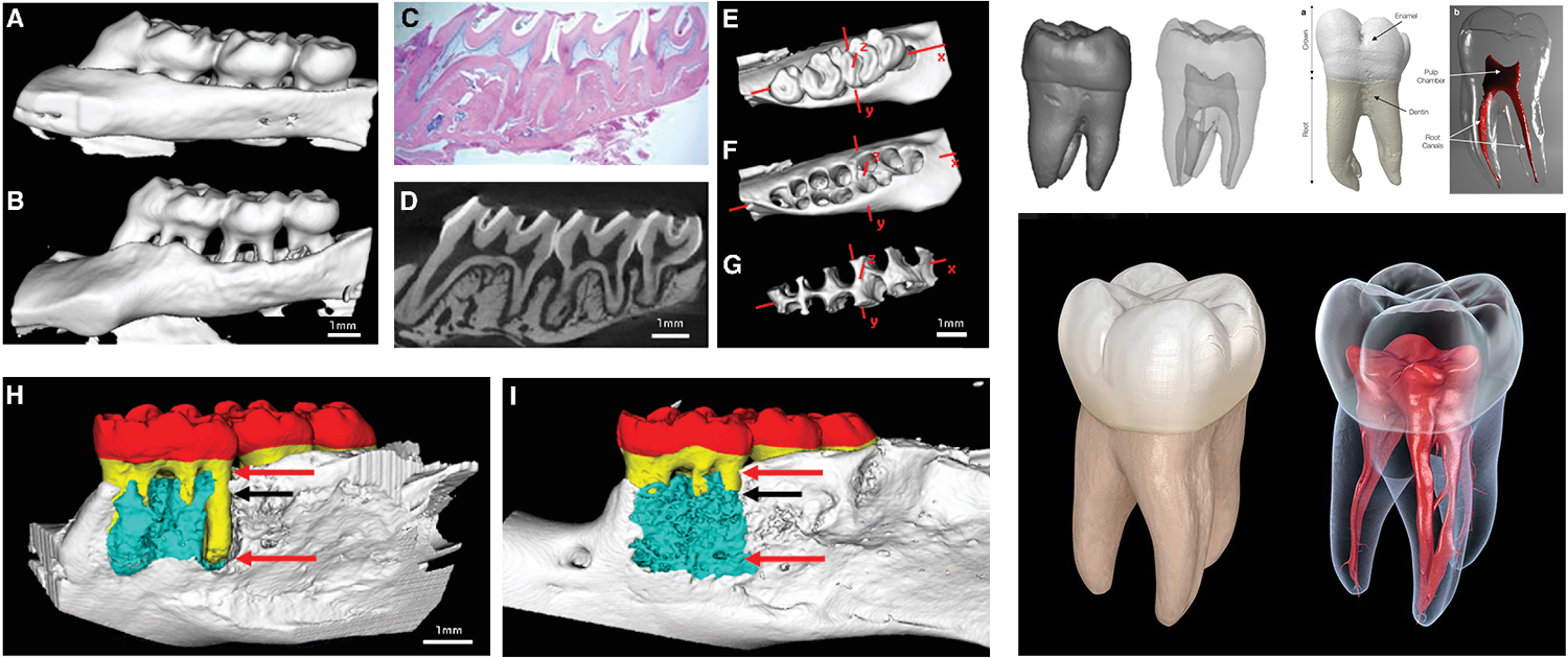
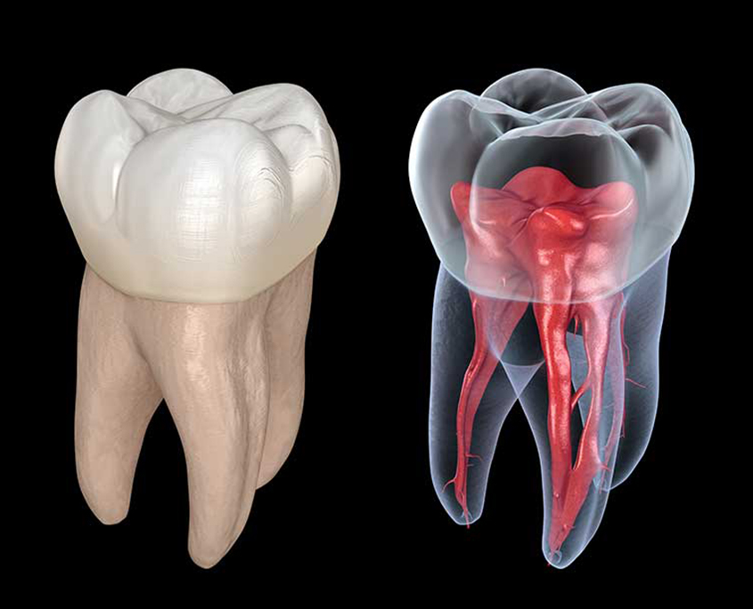
Micro-CT is a powerful tool for studying the anatomy and morphology of the teeth and jaws. It can be used for the study of dental development and eruption, providing important information about the timing and sequence of tooth eruption, as well as the development of the dental arch. Additionally, micro-CT can be used for comparative dental anatomy, providing information about the evolution of teeth and the functional morphology of dental structures in different species.
- Tooth morphology analysis: Micro-CT can be used to analyze the internal and external morphology of teeth, including the roots and pulp cavities. This information is helpful in understanding tooth development and studying dental pathologies.
- Enamel analysis: Micro-CT can be used to analyze the thickness and mineral content of enamel, which is important for understanding the mechanical properties of teeth and for studying enamel-related diseases such as enamel hypoplasia.
- Dentin analysis: Micro-CT can also be used to analyze the structure and density of dentin, which is important for understanding the mechanical properties of teeth and for studying dentin-related diseases such as dentinogenesis imperfect.
Dental material analysis
Micro-CT is an important tool for analyzing dental materials and providing information about the mechanical properties and degradation of different materials used in dentistry. It can also be used to evaluate the osseointegration of dental implants, providing detailed information about bone healing and the success of the implantation process. Finally, micro-CT can be used to assess the accuracy of dental restorations, providing valuable information about the fit and adaptation of different restorative materials.
- Restorative materials analysis: Micro-CT can be used to analyze the internal structure of vital materials such as composites and ceramics, allowing researchers to evaluate their mechanical properties and biocompatibility.
- Implant analysis: Micro-CT can be used to analyze the internal structure of dental implants, allowing researchers to evaluate their osseointegration and long-term stability. This information is essential for developing new implant materials and designs.
- Adhesive interface analysis: Micro-CT can also be used to analyze the interface between dental materials and tooth structure, allowing researchers to evaluate the quality of the bond and develop new bonding agents.
Dental imaging and diagnosis
Micro-CT is a powerful tool for high-resolution dental imaging, providing detailed information about the anatomy and structure of the teeth and jaws. It can be used for quantitatively assessing dental caries, providing important information about the extent of decay and the efficacy of different treatments. Additionally, micro-CT can be used to diagnose endodontic pathologies, such as root canal infections, by visualizing the internal structures of the teeth in 3D.
Orthodontic research
Micro-CT is an important tool for orthodontic research, providing information about the outcomes of orthodontic treatment and the underlying biomechanics of tooth movement. It can be used to evaluate the extent of orthodontic root resorption, providing important information about the risks and benefits of different treatment approaches. Additionally, micro-CT can be used to study the biomechanics of orthodontic tooth movement, providing valuable information about the forces and stresses involved in tooth movement.
Forensic dentistry
Micro-CT is a valuable tool for forensic dentistry, providing detailed information about the teeth and jaws that can be used for dental identification. It can also be used for age estimation, providing information about tooth development and eruption that can be used to estimate the age of an individual. Finally, micro-CT can be used for the study of dental trauma, providing information about the extent and nature of dental injuries, as well as the efficacy of different treatment approaches.
Periodontal Disease Study
Micro CT is an effective tool for studying periodontal disease and its effects on the jaw bones and teeth. Researchers can use these images to better understand the biology of the disease and to develop new and improved treatments.
- Periodontal structure analysis: Micro-CT can be used to analyze the structure of periodontal tissues, including the alveolar bone and periodontal ligament. This information is useful in understanding the biomechanics of teeth and for studying periodontal diseases such as periodontitis.
- Tooth movement analysis: Micro-CT can be used to analyze tooth movement in response to orthodontic forces, allowing researchers to study the biomechanics of orthodontic treatment and to develop new treatment modalities.
- Regenerative medicine analysis: Micro-CT can be used to evaluate the efficacy of regenerative medicine approaches for treating periodontal defects, allowing researchers to develop new therapies for periodontal disease.
Endodontic Treatment Evaluation
Micro CT can be used to evaluate the success of endodontic treatments such as root canal therapy. Researchers can use these images to study the anatomy of the root canals and to assess the effectiveness of the treatment in terms of sealing the canals and preventing reinfection.
- Root canal analysis: Micro-CT can be used to analyze the internal structure of root canals, allowing researchers to evaluate the efficacy of endodontic treatments and develop new treatment modalities.
- Periapical pathology analysis: Micro-CT can be used to analyze periapical pathology, such as apical periodontitis, allowing researchers to study the underlying mechanisms of these conditions and develop new treatment approaches.
- Root resorption analysis: Micro-CT can also be used to analyze root resorption, which is a common complication of orthodontic treatment. This information is useful in understanding the mechanisms of root resorption and in developing new approaches to prevent or treat this condition.
Dental Implant Research
Micro CT can be used to evaluate the stability of dental implants and to assess their integration with the surrounding tissues. This information is useful for researchers developing new and improved implant designs and surgical techniques.
- Implant stability analysis: Micro-CT can be used to analyze the stability of dental implants, allowing researchers to evaluate the efficacy of different implant designs and materials.
- Bone-implant interface analysis: Micro-CT can be used to analyze the interface between bone and dental implants, allowing researchers to study the osseointegration process and to develop new implant materials and designs.
- Implant placement planning: Micro-CT can also be used for implant placement planning, allowing clinicians to evaluate the bone volume and quality at potential implant sites and to plan the optimal implant placement for each patient.
Other Industrial Applications
Quality control and inspection
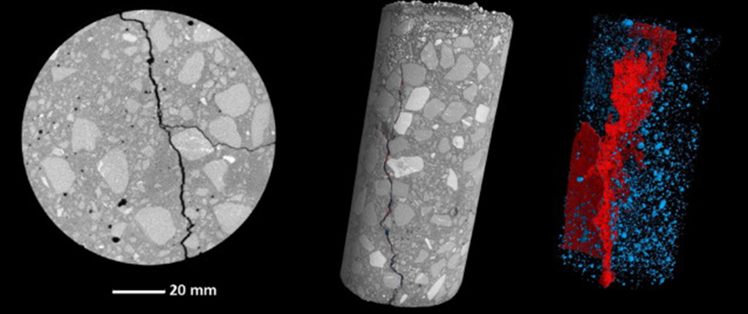
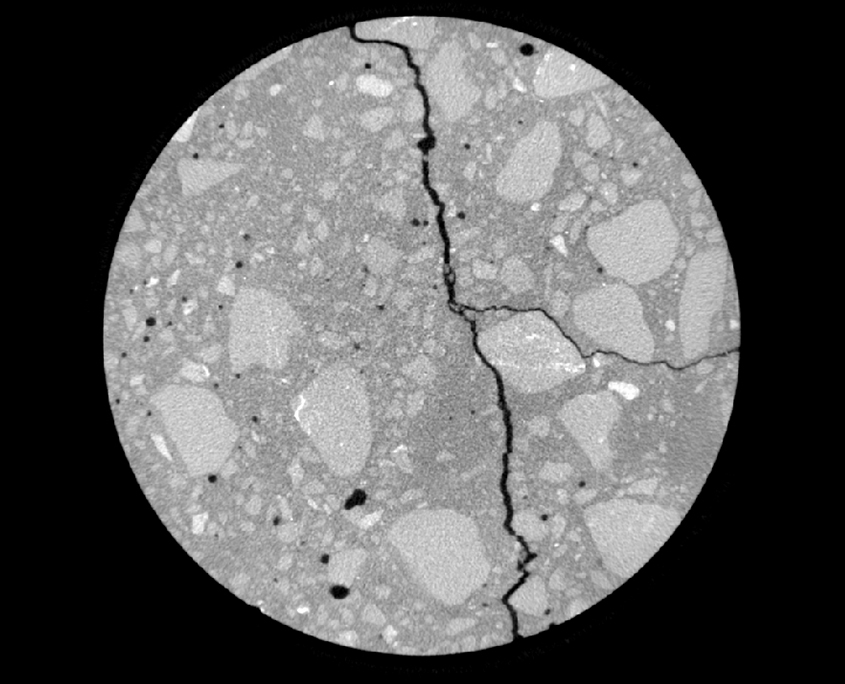
Micro-CT is a valuable tool for quality control and inspection in the industry, providing a non-destructive way to inspect and analyze the internal structure of materials and products. It can be used to detect and quantify defects, such as cracks, voids, and porosity, and to assess the quality of manufactured parts, such as those produced by additive manufacturing.
Materials science and engineering
Micro-CT is an important tool for materials science and engineering, providing detailed information about the microstructure and properties of materials. It can be used to study the microstructure of materials, including their morphology, distribution, and orientation. Additionally, micro-CT can be used to analyze composites and multiphase materials, providing important information about their structure and behavior. Finally, micro-CT can be used to understand the failure mechanisms of materials, providing important insights into how materials break and how they can be improved.
- Porosity and connectivity analysis: Micro-CT can be used to analyze the internal structure of materials, allowing researchers to measure the porosity, connectivity, and morphology of different materials. This information is useful for optimizing the manufacturing process, developing new materials, and evaluating the quality of materials for specific applications.
- Defect analysis: Micro-CT can also be used to detect and characterize defects in materials, including cracks, voids, and inclusions. This information is important for assessing the structural integrity of materials and for developing new testing and quality control methods.
- Failure analysis: Micro-CT can be used to analyze the microstructure of failed components, allowing researchers to identify the root cause of failure and to develop new design and manufacturing processes to prevent future failures.
Geology and earth sciences
Micro-CT is a valuable tool for geology and earth sciences, providing information about the structure and properties of geological materials, such as rocks and minerals. It can be used to quantitatively analyze porosity, providing information about the distribution and connectivity of pores and fractures. Additionally, micro-CT can be used to study the mechanical and physical properties of rocks and minerals, providing important insights into their behavior and potential uses.
Medical devices and engineering
Micro-CT is an important tool for biomedical research and engineering, providing information about the structure and properties of bone and tissue. It can be used to study the microstructure of bone and tissue, providing important insights into their mechanical and biological properties. Additionally, micro-CT can be used to analyze medical devices and implants, providing information about their structure, composition, and performance. Finally, micro-CT can be used to understand disease and injury mechanisms, providing insights into the underlying causes and potential treatments.
- Drug delivery system analysis: Micro-CT can be used to analyze the structure and distribution of drug delivery systems, including micro- and nanoparticles, liposomes, and micelles. This information is important for optimizing the drug delivery process and for developing new drug delivery systems with improved efficacy.
- Medical device analysis: Micro-CT can be used to analyze the internal and external structure of medical devices, including stents, implants
Energy and environmental sciences
Micro-CT is a valuable tool for energy and environmental sciences, providing information about the structure and properties of materials and systems used in energy production and environmental remediation. It can be used to analyze energy materials and devices, providing insights into their structure, performance, and potential improvements. Additionally, micro-CT can be used to study the microstructure of porous media, such as rocks and soils, providing information about their properties and behavior. Finally, micro-CT can be used to understand the transport properties of porous materials, providing insights into how fluids and gases move through these materials.
Additive manufacturing
- Print quality analysis: Micro-CT can be used to analyze the quality of additive manufacturing processes, including layer thickness, porosity, and surface finish. This information is important for optimizing the printing process and for developing new printing materials and techniques.
- Defect analysis: Micro-CT can also be used to detect and characterize defects in printed components, including voids, cracks, and warping. This information is important for evaluating the structural integrity of printed components and for developing new post-processing techniques to improve the quality of printed components.
- Material analysis: Micro-CT can be used to analyze the internal structure and composition of printed materials, allowing researchers to optimize the printing process and to develop new printing materials with improved properties.
Electronics and semiconductor industry
- Package analysis: Micro-CT can be used to analyze the internal structure of electronic packages, including the solder joints, wires, and die. This information is important for assessing the quality of the package and for detecting defects and failure mechanisms.
- Failure analysis: Micro-CT can also be used to analyze the microstructure of failed electronic components, allowing researchers to identify the root cause of failure and develop new design and manufacturing processes to prevent future failures.
- Circuit board analysis: Micro-CT can be used to analyze the internal structure of circuit boards, including the layers, vias, and interconnects. This information is important for optimizing the design and manufacturing process and for detecting defects and failure mechanisms.
Aerospace and automotive industry
- Part analysis: Micro-CT can be used to analyze the internal and external structure of aerospace and automotive parts, including engine components, composite materials, and turbine blades. This information is important for assessing the quality of the part and for detecting defects and failure mechanisms.
- Failure analysis: Micro-CT can also be used to analyze the microstructure of failed aerospace and automotive components, allowing researchers to identify the root cause of failure and to develop new design and manufacturing processes to prevent future failures.
- Corrosion analysis: Micro-CT can be used to analyze the corrosion behavior of aerospace and automotive parts, allowing researchers to evaluate the effectiveness of corrosion prevention methods and to develop new corrosion-resistant materials and coatings.
What can we do for you?
We offer a wide range of preclinical imaging services, including micro-PET, micro-SPECT, micro-CT, and optical imaging. Our team of experienced specialists is dedicated to providing high-quality and reliable image analysis services to help researchers in the preclinical research field. Researchers can register their research projects on our website and choose the services they need or set up a free virtual meeting with our specialists to discuss and get guidance about the available related services that we can perform for them.
Our image analysis services are customizable and can be tailored to fit the specific needs of each research project. With our state-of-the-art equipment and cutting-edge image analysis techniques, we aim to help researchers obtain the most accurate and informative results from their preclinical imaging studies.
To register your research project with our company, please register your project through the “Contact Us” page on our website and fill out the required fields, including your name, email address, and project details.
We will review your submission and contact you as soon as possible to schedule a free virtual meeting with our imaging specialists, who can provide guidance and support for your research.

