
Micro-CT Imaging and Quantitative Analysis of Enamel and Dentin
Enamel and dentin are the primary mineralized tissues of the tooth and play essential roles in protecting the tooth structure and maintaining its mechanical integrity. Understanding their microstructure, mineral distribution, and structural properties is fundamental to dental research, including studies of dental caries, demineralization, remineralization, erosion, abrasion, and the development of new preventive and restorative treatments.
At KARA, we provide high-resolution Micro-CT imaging and quantitative image analysis for enamel and dentin research. Our imaging and analysis workflows enable researchers to investigate the three-dimensional structure of dental hard tissues and quantify changes in mineral density, tissue thickness, lesion characteristics, porosity, and other structural parameters. These approaches can be used to evaluate treatment effects, compare experimental groups, and investigate structural changes in enamel and dentin while preserving the integrity of the samples.
Our services can support a wide range of in vitro and experimental dental research, including studies investigating enamel and dentin microstructure, demineralization and remineralization, dental caries, erosion, preventive treatments, and dental biomaterials.
What Can Be Evaluated?
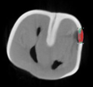
Mineral Density
Micro-CT can be used to evaluate mineral density and its spatial distribution within enamel and dentin. Quantitative assessment of mineral density can help researchers investigate mineral loss, mineral recovery, and differences between experimental groups.
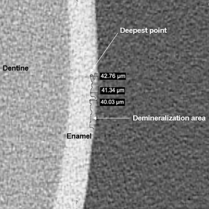
Lesion Depth
Lesion depth can be quantified to assess the extent of structural changes caused by demineralization, dental caries, or other experimental conditions. This parameter is particularly useful for evaluating treatment effects in demineralization and remineralization studies.
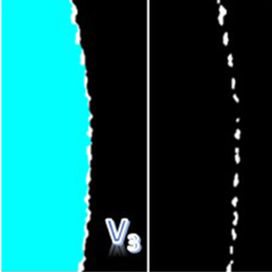
Lesion Volume
Three-dimensional Micro-CT datasets allow the volume of affected or demineralized regions to be quantified. Lesion volume can provide a more comprehensive assessment of structural changes than measurements based on a single two-dimensional section.
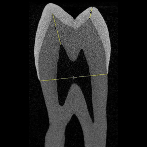
Enamel Thickness
Enamel thickness can be evaluated across different regions of the tooth to characterize its three-dimensional structure and identify regional variations. This parameter can also be used to investigate structural changes associated with experimental treatments or tissue loss.
Dentin Thickness
Quantitative analysis of dentin thickness can provide information about the structural characteristics of dentin and changes associated with experimental conditions. Measurements can be performed in selected regions or across the three-dimensional volume of the sample.
Research Areas
Supporting Research in
- Enamel Microstructure
- Dentin Microstructure
- Dental Caries
- Demineralization & Remineralization
- Dental Erosion
- Preventive Dental Treatments
- Biomimetic Remineralization
- Dental Biomaterials