
Micro-CT Imaging and Quantitative Analysis for Dental Biomaterials Research
The development and evaluation of dental biomaterials require a detailed understanding of their internal structure, morphology, porosity, and interaction with dental tissues. From restorative and filling materials to scaffolds, cements, adhesives, and biomimetic materials, three-dimensional imaging can provide valuable insights into material structure and performance.
At KARA, we provide Micro-CT imaging and quantitative image analysis for dental biomaterials research. High-resolution Micro-CT imaging enables non-destructive three-dimensional visualization of materials and dental structures, allowing researchers to investigate internal morphology and structural changes without sectioning or destroying the samples.
Depending on the material and study design, Micro-CT datasets can be analyzed to evaluate parameters such as material volume, porosity, pore size and distribution, surface area, interfacial gaps, material adaptation, structural integrity, and three-dimensional morphology. These analyses can support the development and evaluation of restorative materials, dental cements, scaffolds, bone substitutes, biomimetic materials, and other innovative dental biomaterials.
Our services can support a wide range of in vitro and experimental dental biomaterials studies, including material characterization, restorative dentistry, endodontic materials, regenerative dentistry, scaffold evaluation, and studies investigating the interaction between dental materials and biological tissues.
What Can Be Evaluated?
Material Volume
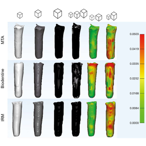
Micro-CT imaging can be used to quantify the three-dimensional volume of dental biomaterials within a sample. Volume measurements can help researchers evaluate material distribution, changes following treatment, and differences between experimental groups.
Porosity
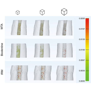
Porosity analysis can provide information about the internal structure of dental biomaterials. Quantifying the total pore volume and porosity can help researchers investigate material structure and compare different formulations or processing methods.
Pore Size and Distribution
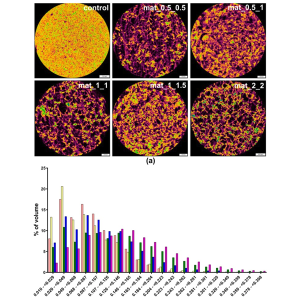
Three-dimensional image analysis can be used to characterize pore size and spatial distribution within porous biomaterials and scaffolds. These parameters are particularly relevant to research involving regenerative dentistry and tissue engineering.
Material–Tissue Interface
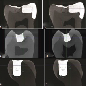
Micro-CT imaging can support the visualization and analysis of the interface between dental materials and surrounding tooth or bone structures. Depending on the material properties and image contrast, interfacial characteristics can be evaluated in three dimensions.
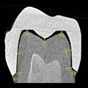
Interfacial Gaps
Quantitative image analysis can be used to investigate gaps or voids at the interface between restorative materials and dental tissues. Three-dimensional analysis may provide information about the volume and spatial distribution of interfacial gaps.
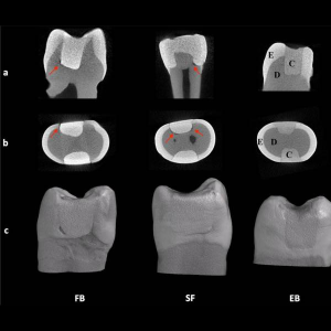
Material Adaptation
Three-dimensional Micro-CT analysis can help researchers evaluate how well a dental material adapts to the surrounding tooth structure. This approach can be useful for investigating the adaptation of restorative materials, cements, and filling materials.
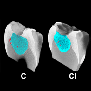
3D Morphology and Structural Integrity
Three-dimensional reconstruction enables visualization of the overall morphology and internal structure of dental biomaterials. Researchers can investigate structural integrity, defects, cracks, and other morphological characteristics when they are detectable at the selected imaging resolution.
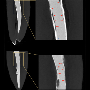
Volume Changes
Micro-CT imaging at multiple time points can be used to investigate changes in material volume or structure over time. This approach may support studies of degradation, dissolution, swelling, shrinkage, or structural changes following experimental treatments.
Research Areas
Supporting Research in
- Dental Restorative Materials
- Dental Cements
- Endodontic Materials
- Dental Adhesives
- Resin-Based Materials
- Biomimetic Materials
- Porous Biomaterials
- Scaffolds for Regenerative Dentistry
- Bone Substitutes
- Tissue Engineering
- Regenerative Dentistry
- Dental Material–Tissue Interaction