
Micro-CT Imaging and Quantitative Analysis for Bone and Periodontal Research
Bone and periodontal research requires detailed evaluation of the complex three-dimensional structure of mineralized tissues and their response to disease, treatment, and regenerative approaches. Understanding changes in bone microarchitecture, alveolar bone loss, periodontal defects, and tissue regeneration is essential for investigating periodontal disease, bone remodeling, and new regenerative therapies.
At KARA, we provide Micro-CT imaging and quantitative image analysis for dental bone and periodontal research. High-resolution Micro-CT enables non-destructive three-dimensional visualization of alveolar bone, jawbone, periodontal defects, and mineralized tissues, allowing researchers to investigate structural changes and treatment outcomes in detail.
Depending on the study design and imaging resolution, Micro-CT datasets can be analyzed to evaluate parameters such as bone volume, bone volume fraction (BV/TV), bone mineral density, trabecular thickness, trabecular number, trabecular separation, cortical bone thickness, bone loss, defect volume, and three-dimensional defect morphology. These measurements can support the evaluation of periodontal disease models, bone remodeling, bone regeneration, graft materials, scaffolds, and regenerative treatments.
Our services support a wide range of in vitro, ex vivo, and preclinical dental and periodontal studies, including periodontal disease research, alveolar bone loss, bone regeneration, guided tissue regeneration, bone graft evaluation, biomaterial and scaffold studies, and the assessment of novel regenerative therapies.
What Can Be Evaluated?
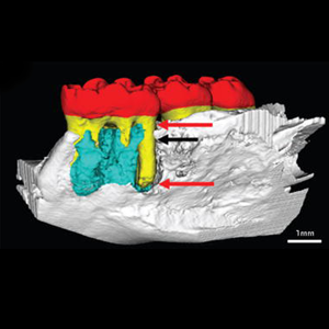
Bone Volume (BV)
Three-dimensional Micro-CT analysis can be used to quantify the volume of mineralized bone within a defined region of interest. Bone volume measurements can help researchers evaluate bone formation, bone loss, and structural changes associated with periodontal disease or regenerative treatments.
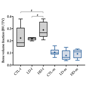
Bone Volume Fraction (BV/TV)
Bone volume fraction represents the proportion of mineralized bone within a defined total volume of interest. BV/TV is widely used to characterize bone quantity and evaluate changes in bone formation, loss, and remodeling.
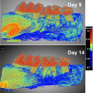
Bone Mineral Density (BMD)
When appropriate calibration and imaging protocols are available, Micro-CT can be used to assess mineral density within bone. Bone mineral density measurements can provide quantitative information about mineralization and changes in bone quality associated with disease or treatment.
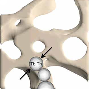
Trabecular Thickness (Tb.Th)
Trabecular thickness provides information about the thickness of trabecular structures within cancellous bone. This parameter can be used to investigate changes in bone microarchitecture associated with periodontal disease, bone loss, or regenerative treatments.
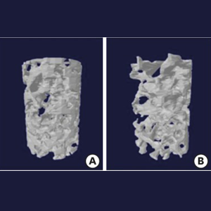
Trabecular Number (Tb.N)
Trabecular number describes the number of trabecular structures within a defined bone volume. Changes in trabecular number can provide information about alterations in bone microarchitecture and the response of bone tissue to disease or regenerative interventions.
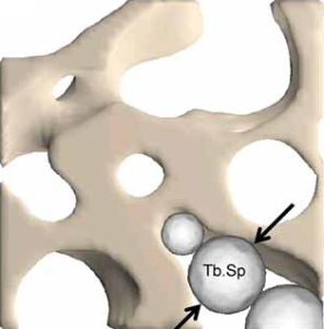
Trabecular Separation (Tb.Sp)
Trabecular separation represents the average distance between trabecular structures. Together with trabecular thickness and number, it can be used to characterize three-dimensional bone microarchitecture and evaluate structural changes over time.
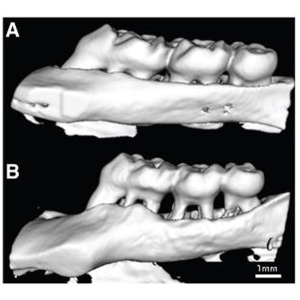
Alveolar Bone Loss
Micro-CT imaging can support quantitative assessment of alveolar bone loss by measuring changes in bone height, volume, or morphology within defined anatomical regions. This approach can be useful for evaluating periodontal disease models and treatment outcomes.
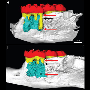
Periodontal and Bone Defect Volume
Three-dimensional segmentation can be used to quantify the volume of periodontal or bone defects. Defect volume measurements can help researchers evaluate the extent of tissue loss and monitor changes following regenerative treatments.
Defect Morphology and 3D Architecture
Three-dimensional reconstruction enables detailed visualization of the shape and spatial characteristics of periodontal and bone defects. This approach can provide comprehensive information about defect morphology and structural changes following treatment.
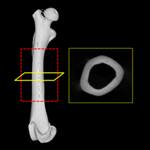
Cortical Bone Thickness
Cortical bone thickness can be evaluated in selected anatomical regions to investigate changes in cortical structure associated with disease, bone remodeling, or regenerative interventions.
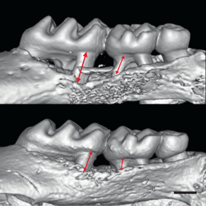
Bone Regeneration
Longitudinal or comparative Micro-CT analysis can help researchers evaluate new mineralized tissue formation within bone defects. Changes in bone volume, mineral density, and defect filling can provide quantitative information about the effectiveness of regenerative treatments.
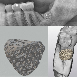
Bone Grafts and Scaffolds
Micro-CT can support the evaluation of bone grafts and scaffolds by visualizing their three-dimensional structure and their relationship with newly formed mineralized tissue. Depending on the material and imaging resolution, quantitative analysis may include scaffold volume, porosity, defect filling, and new bone formation.
Research Areas
Supporting Research in
- Periodontal Disease Research
- Alveolar Bone Loss
- Periodontal Bone Defects
- Bone Microarchitecture
- Bone Remodeling
- Bone Regeneration
- Guided Bone Regeneration (GBR)
- Guided Tissue Regeneration (GTR)
- Bone Grafts
- Bone Substitutes
- Scaffold Evaluation
- Regenerative Dentistry
- Periodontal Regeneration
- Preclinical Periodontal Studies